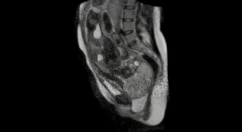
For thousands upon thousands of years, we humans got to witness the blessed spectacle of childbirth from only one vantage point. But now, thanks to the wonders of magnetic resonance imaging technology, or MRI, we can see what’s actually going on.
Putting a soon-to-be mom inside an MRI scanner isn’t some harebrained idea. The point of the experiment, at least when German researchers investigated the possibility in 2010, was to gain a sharper understanding of how the fetus interacts with the pelvis as it travels through the birth canal. The team also wanted to see how various fetal positions could result in spinal complications.
The scans were widely viewed as a success. “Bony structures as well as soft tissue could be assessed in great detail,” the team wrote. Unlike other technologies, which may miss parts of maternal anatomy, MRI was able to capture subtle details like the curve of the uterus that fetuses must navigate. Further research is being designed to expand the technique’s applicability, particularly to stage one of pregnancy. Researchers looked at stage two, right when the baby gets delivered. But stage one examinations can reveal earlier warning signs of arrest of labor, which necessitates C-section.
“This knowledge would allow clinicians to intervene in a timely and effective fashion to ensure a favorable outcome,” the researchers wrote, adding that a basic knowledge of how the fetus’s head is positioned inside the womb “at the time of its passage through the lower part of the birth canal is of practical value in operative vaginal deliveries.”
Leave a Reply